The levels of protein folding are four nested layers: primary sequence, secondary helices and sheets, tertiary whole-chain folding, and quaternary assembly of several folded chains. The layers differ in what holds them together, so no single bond type explains a protein’s shape.
A ribbon diagram on a screen looks like one object, which invites two assumptions: that folding happens in one step, and that one kind of bond holds everything in place. Both are wrong, and the sections below correct them one level at a time. For how structural findings fit into wider research practice, see how do clinical trials work and the other guides in that section.
Misconception one: folding is a single step
Protein structure is organised in layers, and each layer builds on the one below it. The Protein Data Bank’s educational resource PDB-101 states that protein structure is usually broken down into four hierarchical levels of organization, and calls the hierarchy one of the central discoveries about proteins. A chemistry textbook chapter on the same topic names the four levels as primary, secondary, tertiary and quaternary.
The practical consequence is that a question such as “what holds this protein together?” has no single answer. You have to say which level you mean. The table sets the four levels side by side.
| Level | What it describes | Main stabilizing interactions | Example |
|---|---|---|---|
| Primary | The amino acid sequence of one polypeptide chain | Peptide bonds, formed by a dehydration reaction between consecutive amino acids | The residue order of any single chain |
| Secondary | Local folding patterns of the backbone | Hydrogen bonds | Alpha helix and beta pleated sheet |
| Tertiary | Folding of the whole chain, shaped by its side chains | Hydrophobic interactions, ionic bonds, hydrogen bonds and disulfide bridges between side chains | A folded domain of a single chain |
| Quaternary | Two or more folded chains associating into a functional assembly | Association of folded subunits into one complex | Sliding clamp, PDB ID pdb_00006gis, a ring of three subunits |
The bond descriptions come from a study guide on protein structure, folding and misfolding, and the quaternary example comes from the PDB-101 guide to hierarchical structure.
Misconception two: the amino acid sequence is the shape
Primary structure is only the order of amino acids. A University of Vermont course handout defines it as the amino acid sequence of a polypeptide chain, read from the N-terminal end to the C-terminal end. Each amino acid is connected to the next by a peptide bond, and the study guide above adds that these bonds form through a dehydration reaction.
Three points keep this level in perspective:
- Sequence is a linear list. It carries no three-dimensional information on its own. The folded form emerges from the interactions at the higher levels.
- Peptide bonds build the chain, not the fold. They link neighbouring residues. The hydrogen bonds, ionic bonds and hydrophobic interactions that shape the fold act at the secondary and tertiary levels.
- Direction matters. The N-to-C reading order means a chain has a defined start and end, so two chains with the same residues in a different order are different proteins.
That is why sequence is the starting point of every structural description, but never the whole description.
Misconception three: helices and sheets are the finished protein
Secondary structure is local, not global. The University of Vermont handout describes it as the local spatial arrangement of a polypeptide’s backbone (main chain) atoms, and the study guide notes that hydrogen bonds stabilize both the alpha helix and the beta pleated sheet. The side chains do not define this level; the backbone does.
Alpha helix
An alpha helix is an element of secondary structure in which the amino acid chain is arranged in a spiral. According to the Vermont handout, it is right-handed, repeats every 3.6 residues per turn, and rises about 5.4 Å per turn. The hydrogen-bond pattern is regular: the C=O of residue i bonds to the NH of residue i+4. PDB-101 gives a more visual description, saying alpha helices form spring-shaped structures with each amino acid hydrogen bonding to one further down the chain.
Beta strands and sheets
Adjacent beta strands can hydrogen bond to one another to form a beta sheet, also called a beta pleated sheet. The bonding here runs between neighbouring strands rather than along a single stretch of chain, which is the main contrast with the helix.
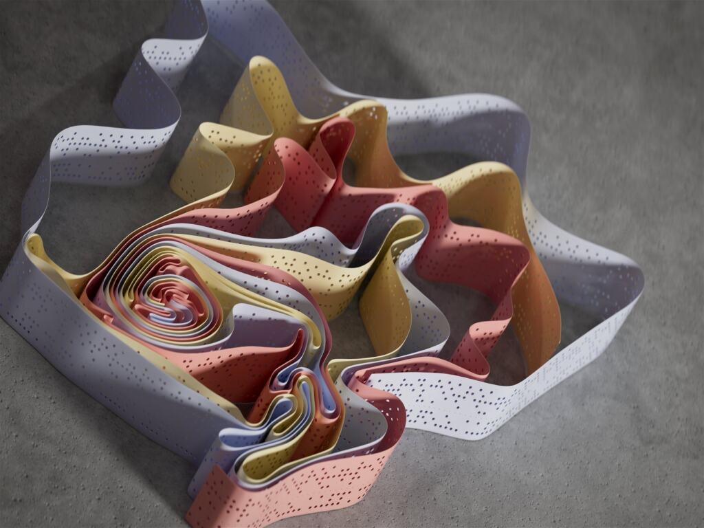
Helices and sheets are building blocks. They have to be arranged relative to each other, and that arrangement is the next level.
Misconception four: the backbone hydrogen bonds hold the whole fold
Tertiary structure is mainly a story about side chains. The SWISS-MODEL course chapter describes it as the folding of the polypeptide chain to assemble secondary structure elements into a three-dimensional arrangement. The study guide specifies that it is formed by interactions among side chains (R groups): hydrophobic interactions, ionic bonds, hydrogen bonds and disulfide bridges.
Several practical readings follow from that list:
- Mixed forces, not one. A tertiary fold is the sum of several interaction types, so changing a single residue can matter differently depending on which interaction it contributed to.
- Helices pack together. SWISS-MODEL gives the globin family as an example, with eight alpha-helices in contact. A tertiary fold can be built almost entirely from one kind of secondary element.
- Domains are the unit. SWISS-MODEL says that as helices and sheets are units of secondary structure, the domain is the unit of tertiary structure. A chain can therefore contain more than one domain.
If you are trying to understand why a mutation changes a protein, look first at which side-chain interactions the changed residue took part in. The backbone pattern alone will rarely explain it.
Misconception five: every protein has quaternary structure
Quaternary structure exists only when separate folded chains come together. PDB-101 defines it as two or more folded chains associating to form a functional assembly. A protein that works as a single folded chain is fully described by the first three levels.

Where assemblies do exist, the subunits may be of one type or several. PDB-101 labels identical subunits homo-oligomers and mixed ones hetero-oligomers.
A worked PDB example: the sliding clamp
The clearest case in the PDB-101 guide is the sliding clamp protein, PDB ID pdb_00006gis. It forms a ring that surrounds DNA, and three folded subunits associate to make that ring-shaped quaternary structure. Each subunit is itself a folded chain with its own tertiary structure, so the ring only exists at the quaternary level. Looking up that entry in the Protein Data Bank lets you see the levels stacked: the sequence, the helices and sheets, the folded subunits, and the assembled ring.
Misconception six: a disrupted protein has simply broken apart
Disruption mostly undoes the higher levels. The study guide says external factors such as heat, pH changes or chemicals can disrupt protein structure, a process called denaturation, and that it involves disruption of non-covalent interactions: hydrogen bonds, ionic bonds and hydrophobic interactions. Those are the interactions that stabilize secondary, tertiary and, by extension, assembled structures, which is why loss of shape tends to mean loss of function.
The same guide links misfolding to disease. It characterizes Alzheimer’s disease by amyloid plaques formed from misfolded proteins in the brain. The lesson for reading the literature is that a protein can have a perfectly correct sequence and still fail, because the fold, not the sequence alone, decides whether it works.
Where to go next
Structure is one layer of how cells control what proteins do. For regulation that acts above the sequence itself, read What Is Epigenetics? How Genes Are Switched On and Off — A Clear Guide For 2026 – Biolifetechnologies. For a hands-on laboratory context, see How To Make Organoids: A Practical Lab Guide For Beginners (2026) – Biolifetechnologies.
Frequently asked questions
| Question | Answer |
|---|---|
| How much of a typical protein is beta strand? | The University of Vermont handout says approximately a quarter of all residues in a typical protein are in beta strands. The rest is in other secondary structure and connecting regions. |
| Can a quaternary assembly contain different chains? | Yes. PDB-101 says the individual proteins may be identical (homo-oligomers) or of many different types (hetero-oligomers). |
| What is a domain? | A domain is the unit of tertiary structure, just as helices and sheets are units of secondary structure. SWISS-MODEL notes that tertiary structure includes multi-domain arrangements. |